What is AMD?
Macular degeneration, a disease that affects the center of the retina called the macula, is the leading cause of blindness in adults over the age of 55. It involves a weakening of the retinal tissues that cause gradual vision loss and may develop as a result of genetics, age, diet, smoking and sun exposure. Patients with this condition may experience distorted or blurry vision, a loss of color vision or a dark area in the center of vision. The resulting degeneration can lead to severe central (not peripheral vision) vision loss.
There are two types of macular degeneration: dry and wet. Although the wet variety can more commonly lead to severe visual loss, it is also the type that has more effective treatment options available. Although there is no cure for macular degeneration, early detection and treatment may slow progression of the disease. With the latest in high-resolution digital imaging, we are able to closely follow any progressive degeneration and recommend the latest in treatment options. Regular eye exams can identify whether you are at risk of macular degeneration.
The Macula
The macula is made up of millions of light-sensing cells that provide sharp, central vision. It is the most sensitive part of the retina, which is located at the back of the eye. The retina turns light into electrical signals and then sends these electrical signals through the optic nerve to the brain, where they are translated into the images we see. When the macula is damaged, the center of your field of view may appear blurry, distorted, or dark.
Who is at risk?
Age is a major risk factor for AMD. The disease is most likely to occur after age 60, but it can occur earlier. Other risk factors for AMD include:
- Smoking. Research shows that smoking doubles the risk of AMD.
- Race. AMD is more common among Caucasians than among African-Americans or Hispanics/Latinos.
- Family history and Genetics. People with a family history of AMD are at higher risk. At last count, researchers had identified nearly 20 genes that can affect the risk of developing AMD. Many more genetic risk factors are suspected.
You may see offers for genetic testing for AMD. Because AMD is influenced by so many genes plus environmental factors such as smoking and nutrition, there are currently no genetic tests that can diagnose AMD, or predict with certainty who will develop it.
The American Academy of Ophthalmology currently recommends against routine genetic testing for AMD, and insurance generally does not cover such testing.
Does lifestyle make a difference?
Researchers have found links between AMD and some lifestyle choices, such as smoking. You might be able to reduce your risk of AMD or slow its progression by making these healthy choices:
- Avoid smoking
- Exercise regularly
- Maintain normal blood pressure and cholesterol levels
Eat a healthy diet rich in green, leafy vegetables and fish
Age-related macular degeneration (AMD) is a deterioration or breakdown of the eye’s macula. The macula is a small area in the retina — the light-sensitive tissue lining the back of the eye. The macula is the part of the retina that is responsible for your central vision, allowing you to see fine details clearly.
The macula makes up only a small part of the retina, yet it is much more sensitive to detail than the rest of the retina (called the peripheral retina). The macula is what allows you to thread a needle, read small print, and read street signs. The peripheral retina gives you side (or peripheral) vision. If someone is standing off to one side of your vision, your peripheral retina helps you know that person is there by allowing you to see their general shape.
Many older people develop macular degeneration as part of the body’s natural aging process. There are different kinds of macular problems, but the most common is age-related macular degeneration.
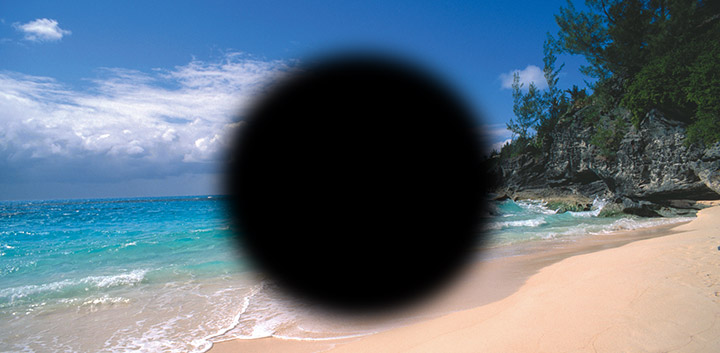
With macular degeneration, you may have symptoms such as blurriness, dark areas or distortion in your central vision, and perhaps permanent loss of your central vision. It usually does not affect your side, or peripheral vision. For example, with advanced macular degeneration, you could see the outline of a clock, yet may not be able to see the hands of the clock to tell what time it is.
Causes of macular degeneration include the formation of deposits called drusen under the retina, and in some cases, the growth of abnormal blood vessels under the retina. With or without treatment, macular degeneration alone almost never causes total blindness. People with more advanced cases of macular degeneration continue to have useful vision using their side, or peripheral vision.
When macular degeneration does lead to loss of vision, it usually begins in just one eye, though it may affect the other eye later.
Many people are not aware that they have macular degeneration until they have a noticeable vision problem or until it is detected during an eye examination.
Types of Macular Degeneration:
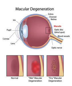
“Dry” Macular Degeneration
Most people who have macular degeneration have the dry form. This condition is caused by aging and thinning of the tissues of the macula. Macular degeneration usually begins when tiny yellow or white pieces of fatty protein called drusen form under the retina. Eventually, the macula may become thinner and stop working properly.
With dry macular degeneration, vision loss is usually gradual. People who develop dry macular degeneration must carefully and constantly monitor their central vision. If you notice any changes in your vision, you should tell your ophthalmologist right away, as the dry form can change into the more damaging form of macular degeneration called wet (exudative) macular degeneration. While there is no medication or treatment for dry macular degeneration, some people may benefit from a vitamin therapy regimen for dry macular degeneration.
Using an Amsler grid to test for macular degeneration
If you have been diagnosed with dry macular degeneration, you should use a chart called an Amsler grid every day to monitor your vision, as dry macular degeneration can change into the more damaging wet form.
To use the Amsler grid, wear your reading glasses and hold the grid 12 to 15 inches away from your face in good light.
- Cover one eye.
- Look directly at the center dot with the uncovered eye and keep your eye focused on it.
- While looking directly at the center dot, note whether all lines of the grid are straight or if any areas are distorted, blurry or dark.
- Repeat this procedure with the other eye.
- If any area of the grid looks wavy, blurred or dark, contact your ophthalmologist.
- If you detect any changes when looking at the grid, you should notify your ophthalmologist immediately.
“Wet” Macular Degeneration
About 10 percent of people who have macular degeneration have the wet form, but it can cause more damage to your central or detail vision than the dry form. Wet macular degeneration occurs when abnormal blood vessels begin to grow underneath the retina. This blood vessel growth is called choroidal neovascularization (CNV) because these vessels grow from the layer under the retina called the choroid. These new blood vessels may leak fluid or blood, blurring or distorting central vision. Vision loss from this form of macular degeneration may be faster and more noticeable than that from dry macular degeneration.
The longer these abnormal vessels leak or grow, the more risk you have of losing more of your detailed vision. Also, if abnormal blood vessel growth happens in one eye, there is a risk that it will occur in the other eye. The earlier that wet macular degeneration is diagnosed and treated, the better chance you have of preserving some or much of your central vision. That is why it is so important that you and your ophthalmologist monitor your vision in each eye carefully.
Treatment for wet macular degeneration involves the injection of medications into the center of the eye. These medications act on the abnormal growth of blood vessels in the macula that cause wet macular degeneration.
Adapt DX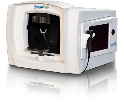
Adapt DX is the newest techonology available for the early detection of macular degeneration. In its earliest form, macular degeneration begins with small, focal changes in the deep layers of the retina. These changes are called “drusen”. Adapt DX allows us to identify patients at an earlier stage in order to begin vitamin therapy, as
well as make healthy lifestyle recommendations. This technology uses dark adaptation testing in order to identify at-risk patients. It is simple and painless, and can be completed typically in less than 10 minutes. We are excited to bring this technology to Pittsburgh.